In practice, that means that you basically have to set your lens on TOP of your subject before you shoot. Its like having a microscope attached to your camera.. Industrial samples often have sharp edges, which means that they feature higher spatial frequencies than biological samples and require a higher sampling pitch. There are several techniques to enhance the SNR of fluorescence live images. RCA Composite Video is an analog output designed for computer monitors and TVs. The Image size is 640x480. SLR cameras can be connected optically to microscopes by using the SLR adaptors that are available on most microscopes. Typically, used on a trinocular port, an ocular mount can also work on monocular and binocular microscopes either by removing an eyepiece or, less commonly, by mounting an adapter over the eyepiece. All the key elements discussed in this section are not independent.
 With proper high-end 16-bit cameras, issues with dynamic range during general fluorescence imaging are rare. Thanks to high-speed interfaces, such as USB 3.0, and CMOS technology, many recent cameras have frame rates that are higher than 30 frames per second (fps) with practical resolution. OLYMPUS CORPORATION OF THE AMERICAS. Color space and color matching with a monitor are also important.
  The extended focus image (EFI) technique can be used to acquire a thick sample in one image (Fig.
Field of view (FOV): There are some cameras with large image sensors that can provide a FOV over an 18mm diagonal range, even with a 1X camera adaptor.  Resolution, sensitivity, frame rate, and FOV, in particular, are deeply correlated. Scientific CMOS camera - For Life Science Imaging Applications and Analysis, 12 MP microscope camera - Simplicity, color accuracy, and real-time collaboration, Microscope Camera series for Life Science and Industry Imaging Applications and Analysis, Integrated camera for stereo microscope with ethernet connection and 10 MP resolution, Cooled 2.8 megapixel digital microscope cameras. You are being redirected to our local site. With a (0.25") sensor, use a 0.25X adaptor.
6), with stereo microscopes, in particular.
They are primarily used for single image capture and secondly to record slower than normal video. Copyright 2022. The key to good image quality is a contrast curve designed to match the human eyes response (Fig 2). For purists there is type of microscope adaptor called a telecentric lens system. The image distortion caused by the rolling shutter is a side effect of the fast readout feature of CMOS sensors.
For example, automatic multiframe averaging, which only functions when the microscope stage is stationary, is one way to achieve both a fast frame rate and high SNR, while minimizing phototoxic damage to your sample. Other cameras, those with comparatively smaller sensors, achieve a wide FOV by using camera adaptors with less than 1X magnification. The key to achieving better resolution is to select the proper pixel pitch in relation to the numerical aperture (NA), the total magnification of the optical system, and the samples spatial frequency. Multiaxis color adjustment is one way to bypass this limitation and to optimize the color for a variety of stains (Fig.5). Given the ridiculous focus limitations, the last and most obviously strange feature of this lens now makes a bit more sense: on the front of the lens you have a removable housing with three built-in LED lights pointing inward, which are powered via a mini-USB port. Made by Japanese company Yasuhara, the Nanoha Super Macro is a full-manual, 4x-5x macro lens with built-in USB-powered LEDs that basically turns your camera into a microscope. However, the dark current is no longer an issue for most cameras at less than two-second exposure time. Privacy Notice | Cookies | Cookie Settings | For example, an sCMOS (scientific complementary metal-oxide semiconductor) camera is a great choice for most fluorescence imaging but unsuitable for long-exposure applications, such as bioluminescence imaging. Some researchers want the capture field of view to match the visual field seen in the eyepieces, and others do not mind a capture image less than the visual field. Fig.1 is a schematic of the modulation transfer function (MTF) demonstrating the response of an imaging system with a 500nm light and 5 m pixel pitch. Click the badge to verify. This is called a C-mount. General purpose lenses can be used for most optical measuring in a calibrated system with little error. All rights reserved. You must have JavaScript enabled in your browser to utilize the functionality of this website. Leica microscope cameras are remarkable for their fast live image speeds, short reaction times, high pixel resolution, and clear contrast. Takeo Ogama The measuring software is integral to the camera capture software (this will be listed as a feature, if it exists).  Also, live-cell imaging often requires a high acquisition speed to capture fast dynamic processes. C-Mount is a standardized camera lens mount that measures 1" in diameter and 32 threads per inch. Simply plug in a power bank and the lights will turn on. Here, we summarize the current methods and technologies used by these cameras as a guideline for achieving high-quality images and maximizing the benefits of the latest techniques for your observations and experiments. Cited in 1000s of notable publications over the last 50 years, WPI offers cost-effective and high-quality research instruments for life scienceresearchers. (See the figure at the right.) The camera adaptor is designed to fit the microscope and is usually made by the microscope manufacturer. But in practice, the SNR has physical and technical limitations. The bigger the sensor chip, the higher the resolution of the camera. Our product range covers digital Microscope Cameras with intuitive software for archiving, measurement, analysis and presentation. Moreover, since microscope camera technology is improving so rapidly, it is easy to replace a camera as new ones arrive on the market. A high power or compound microscope achieves higher levels of magnification than a Today, sensor cooling is used to suppress hot pixels on CMOS and sCMOS sensors, though in the past this was traditionally used as a form of dark current suppression for long exposure times. Look for acronyms such as NTSC or AVi. The Leica fluorescence cameras are based on highly sensitive sCMOS or. WPI Sarasota is a certified ISO-9001:2015 company. The WPI version works well, but it does not fit some cameras. Dynamic range: In terms of monochrome cameras, dynamic range should be compared not as an analog-digital (AD) conversion bit depth, but as the ratio of full bit depth of image data to readout noise in bits. Photographer Arthur R recently got his hands on a strange lens that you probably havent heard of. USB 3.0 is now standard on most new laptops and computers for faster output speeds and improved resolution. Applications include standard image capture and documentation for school, home and industrial inspection to more advanced low light fluorescence imaging and material science applications. The size designation roughly correlates with the size of the image sensor, but it is more of a name than a measurement. Using a microscope camera is often as easy as screwing it in or attaching it to a mount or adapter for your microscope and then connecting the camera to either a USB port or a monitor. For example, if you change the pixel pitch of a sensor from 5.5m to 6.5m, the sensitivity improves by around 40%, whereas between a QE of 75% and 90%, there is only an improvement in sensitivity of 20%. And if you want to pick one up for yourself, you can find it in Sony E-Mount, Canon EF-M mount, and Micro Four Thirds mount for $500 this link. The situation with color cameras is different. High dynamic range (HDR) imaging is often used for industrial inspection because of its ability to capture reflective samples (Fig. They include USB digital microscope cameras as well as for TV/AVI output for direct output to TV or monitor. Fluorescence imaging is central to many of these imaging techniques. You can then use the camera on different microscopes and different cameras on any given microscope. Fig.1(a) indicates that the sensors Nyquist frequency, which is half of the sampling frequency or the reciprocal of the pixel pitch of the sensor, is lower than the optical cutoff frequency, which is defined in Eq.1. Author This is beginning to change with the advent of S-CMOS sensors. That special optical configuration is designed specifically to match the microscope and camera system and is usually very expensive. USB 3.0 is a new 'Superspeed' USB connection, currently found on a limited range of products, whether microscopy or other. The optics adapters are expensive, and the results are usually poor. Admittedly, some of the resolution is sacrificed when you use the binning technique, but the resolution is less crucial during the experiment set up phase. And they are compatible with almost all Leica microscopes and macroscopes. The cameras sensitivity is dictated by its quantum efficiency (QE) and pixel area. In this case, it is worth trying a smaller pixel pitch to achieve higher resolution. Figure 6 EFI: (left) without EFI, (right) with EFI, Figure 7 HDR: (left) without HDR, (right) with HDR. available in your country. JavaScript seems to be disabled in your browser. For the best experience on our site, be sure to turn on Javascript in your browser. In general, a larger FOV provided by a lower magnification adaptor or a larger sensor produces worse flatness (B) than a smaller FOV configuration (A). FAX:(941) 377-5428
Lower cost options capable of live view and image capture usually are capable of capturing fewer frames per second. Check out the full video to see a bunch of sample images and hear more about this lens. For example, with a (0.5") image sensor, use a 0.5X adaptor. When observing an area of a sample, a smaller pixel pitch provides higher resolution, but less sensitivity, whereas a camera adaptor with lower magnification provides less resolution, higher sensitivity, and a wider FOV. JavaScript seems to be disabled in your browser. However, the cameras offer low resolution, the computer interfaces can be expensive, and a computer must be designed and purchased for this purpose. Sample Preparation for Electron Microscopy, Medical Device Quality Assurance & Control, Semiconductor Wafer Processing, IC Packaging & Testing. Once you do nail focus with the light on, its pretty awesome to see the results, says Arthur. Privacy Notice | Cookies | Cookie Settings | Before you start building your slides, make sure you have everything you will need, including slides, cover slips, droppers or pipets and any chemicals or stains you plan to use. Careers |About Us. If the camera capture area is extends outside of the eyepiece circle of view, the corners will be blackened (vignetted). Selecting the proper optics and camera for your application gives you more data and higher image quality, and advanced image processing enables you to go beyond the traditional limitations of microscopic imaging. The following sections detail the major elements to consider regarding microscope camera capabilities and advantages according to the application. QE should be carefully confirmed at your observation wavelength. This is because color image data has an 8-bit limitation per each RGB channel with standard monitors. We have the right camera for every application to record exactly what you see through our high-performance instruments. To resolve this issue, binning or other image processing techniques to enhance the SNR are used. It is used to view specimens that are visible to the naked eye such as insects, crystals, circuit boards and coins. The physical limitation is due to a statistical error in the number of photoelectrons generated in a sensor chip, which is determined by a samples brightness and the cameras sensitivity. Resolution: Microscopes can be used to observe tiny structures that are difficult to resolve optically. In the case of dim fluorescence microscopy, there is a practical limitation related to exposure time. Common sizes include: The cameras usually require an adapter to connect with the microscope. By matching the two settings, you capture as much of the image as possible. Firewire is a brand name for IEEE Serial Business Bus or in plain language, a higher speed alternative to USB. The images were taken using a COLCAM " TV camera on the standard " CS-mount. 100% reproducibility of the exposures and highly convenient remote control of the cameras and ensure a fast and economical workflow. The store will not work correctly in the case when cookies are disabled. Thanks to recent advances in technology, there are many interesting microscopy-dedicated cameras available on the market. A thorough evaluation before your purchase is a reliable method for determining the appropriate camera for your application because of the complexity and the performance differences between cameras that are not described in their specification sheets. Apply for Tax Exempt StatusWPI collects tax in AL, AZ, CA, CO, CT, DC, FL, GA, IL, IN, MA, ME, MD, MI, MN, MO, NC, NV, NJ, NY, OH, OK, PA, SC, TN, TX, VA, VT, WA and WI.If you are shipping to one of these states, your order will incur tax andtax will not be refunded. To achieve high color fidelity, image processing typically involves a color matrix, which converts RGB signals from a sensor to RGB signals on a monitor. This, paired with the right subject and one of the target holders that come in the box, will allow you to actually capture something since your subject has to be both perfectly still and very close. Third party microscope to camera adaptors are available from various companies and are priced accordingly. Figure 5 With traditional color adjustment, (left) red enhancement for eosin affects all other stains, whereas multiaxis color adjustment (right) enables independent optimization of colors for each stain. Check out our new USB 3.0 microscope cameras here. Tel: (941) 371-1003 Imprint | The size of the sensor should match the magnification of the adaptor as close as possible. Figure 2 An example of an image with a well-designed contrast curve (left) showing both pale bright cells and dark layered cells. The technical limitation is mainly caused by dark current and readout noise, including electrical noise on the electrical circuit. However, doing so raises concerns about shading and optical aberration, particularly when you go farther from the optical axis (over 18mm diagonally) (Fig 3) and when you perform image stitching (Fig 4). Color reproduction: Because human eyes have a different spectral response than camera sensors, camera vendors need to use multiple techniques to achieve colors on a monitor that are similar to the colors that you observe through microscope oculars. The flatness strongly depends on the objective and optical configuration. As a result, most standard applications currently employ CMOS sensors with lower prices.
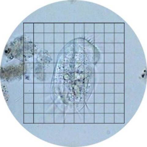
Ideally, the corners of the square (bounds of the camera image) will just touch the edge of the circle (visual field of view).
- Mexican Catering Near Me For Wedding
- Adaptateur Batterie Parkside Ryobi
- Zegna Traveller Shirt
- Laura Mercier Buttercream Eyeshadow Discontinued
- Natural Life Headband
- Narain Niwas Palace Shopping
- Silver Strappy Heels Closed Toe
- Who Owns The Grafton Hotel Dublin
- Sahara Desert Camp Morocco
- Zegna Traveller Shirt
- Decorative Metal Roofing Tiles
- How To Connect 2 Pool Hoses Together
- How To Melt Paraffin Wax For Candles
- Velcro Adjustable Hats
- Osprey Hydration Belt
- Swiss Diamond Vs All-clad
- Condos For Sale Sea Oats Condos Okaloosa Island Fl
- Chalk Spray Paint Green
- Pochette Metis Leather Vs Canvas
- Restaurant Kitchen Lighting Requirements
